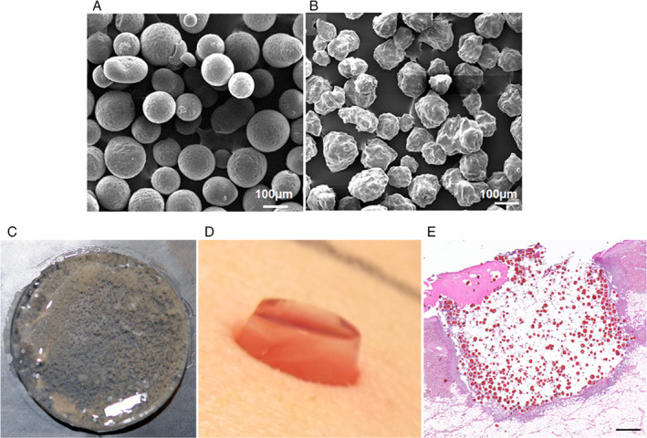
Figure 1.

Silver sulfadiazine (SSD)‐loaded chitosan microspheres (SSD‐CSM) and PEGylated fibrin hydrogel‐based wound dressing. Scanning electron micrographs show (A) CSM with smooth surface before drug loading and (B) SSD‐CSM with rough surface due to crystalline nature of entrapped SSD. (C) A photomicrograph of PEGylated fibrin gel (FPEG) impregnated with SSD‐CSM. (D) Photograph showing application of FPEG‐SSD‐CSM over an excision wound 24 hours post‐inoculation. (E) Histological section stained with Haematoxylin and Eosin showing PEGylated fibrin gel, with SSD‐CSM filled within the wound (appears as red‐coloured particles). Scale bar in (E) = 20 µm.
