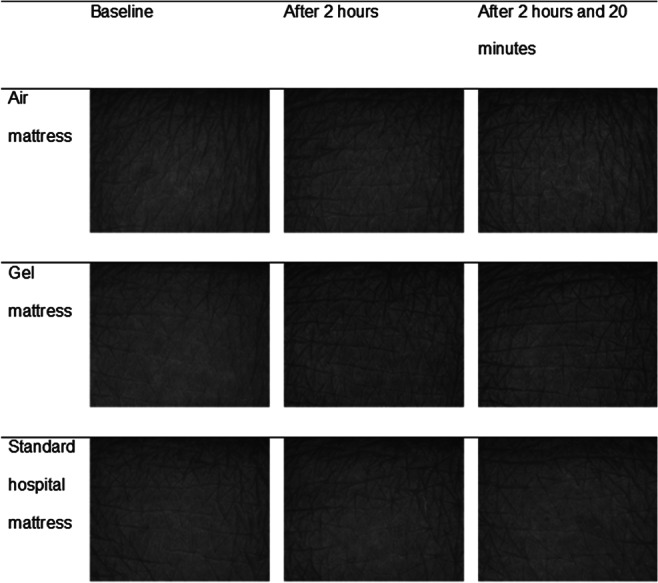
Figure 2.

Visioscan images at sacral skin per intervention and time point for 1 subject. This figure shows skin surface images of the sacral skin taken with Visioscan VC 98 camera before loading (baseline), after 2 hours loading, and 20 minutes after off‐loading per 3 different support surfaces (air, gel, and standard foam)
