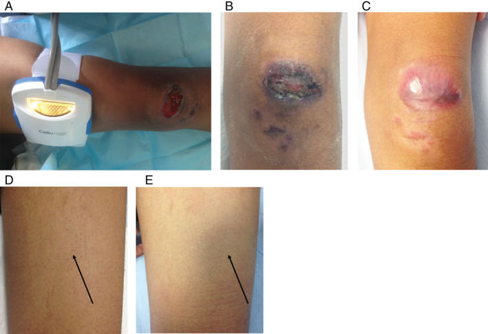
Figure 3.

The wound and donor site of Patient 2/HB. (A) Healthy granulation tissue was seen on the wound bed after 4 weeks of negative pressure wound therapy (NPWT). The wound measures 4·5 × 3·0 cm over the left patella region. (B) More than 50% of the wound was reepithelialised at week 3 postgrafting. (C) Complete wound healing was seen at week 5. (D) Minimal scabs were seen at the donor site (black arrow) at week 3. (E) No visible scar was seen at the donor site (black arrow) at week 6. The donor site looks aesthetically similar to the surrounding skin.
