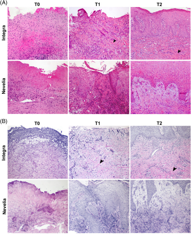
Figure 9.

(A, B) Microscopic aspects of wound healing after Integra and Nevelia application. (A) Representative microscopic images of haematoxylin and eosin‐stained paraffin sections of skin biopsies at baseline (T0) showing typical wounds with cellular debris and dermal inflammatory infiltrate. After the DS application at T1 (after 2 weeks) and at T2 (after 3 weeks), skin punch biopsies showed wound healing with reepithelialisation and dermal granulation tissue with (B) collagen and elastic fibre deposition, as shown by Verhoeff‐Van Gieson staining. Presence of Integra is still evident in dermal tissue at T1 and T2 (arrowheads, original magnification: ×100)
