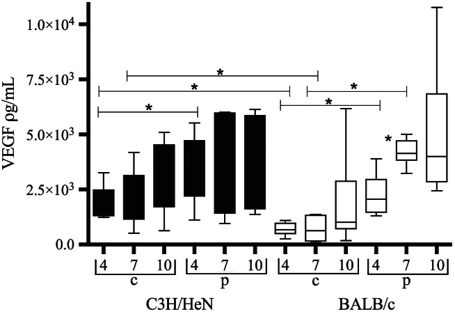
Figure 1.

Vascular endothelial growth factor (VEGF) levels in central (c) and peripheral (p) wound biopsies from infected C3H/HeN mice (black bars) and infected BALB/c mice (white bars). Data are shown as box‐and‐whisker diagrams. VEGF levels are lower centrally than peripherally in BALB/c wounds at days 4 and 7 after infection (P < 0·004; P < 0·0001). In C3H/HeN mice, levels are lower centrally only at day 4 after infection (P < 0·05). In BALB/c mice, levels increase peripherally from 4 to 7 days after infection (P < 0·0018). Comparing the two strains of mice, central VEGF levels are reduced in BALB/c mice day 4 and 7 after infection as compared to C3H/HeN (P < 0·003; P < 0·03). Peripheral levels are equal.
