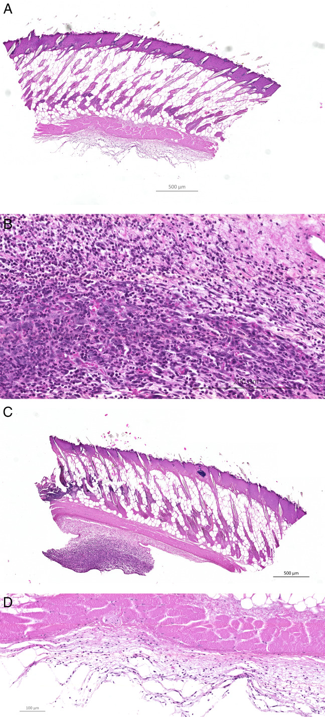
Figure 2.

A representative display of a haematoxylin and eosin (HE)‐stained peripheral (A, B) and central (C, D) wound biopsy taken 10 DPI from the same infected BALB/c mouse (10× and magnification 40×, respectively). Significantly more of the peripheral biopsies taken from infected BALB/c mice had PMN‐dominated inflammation peripherally than central biopsies at 10DPI (P < 0·05).
