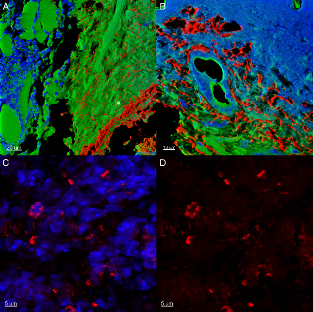
Figure 5.

A representative section of a peripheral biopsy from a Pseudomonas aeruginosa biofilm‐infected (red colour) C3H/HeN mouse at day 7 days post‐infection (DPI). Green colour, local wound tissue; blue colour, inflammatory infiltrate. (A) Scale bar = 20 μm. (B) Scale bar = 10 μm. (C) The appearance of biofilms and inflammatory cells in clusters. Scale bar = 5 μm. (D) The appearance of biofilm in clusters. Scale bar = 5 μm.
