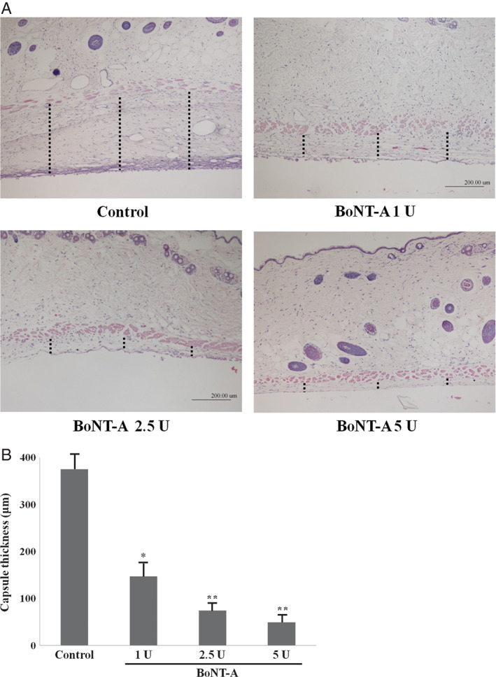
Figure 1.

Representative histological sections of capsular tissues in experimental groups or control group at the 30‐day time point (original magnification, ×40). Sections were stained with haematoxylin and eosin. Length of dot line is capsular thickness. The capsules of experimental groups were significantly thinner than those of control group (P < 0·05). The capsules of experimental group 1 were significantly thicker than those of experimental groups 2 and 3 (P < 0·05). *P < 0·05 versus control; **P < 0·01 versus control.
