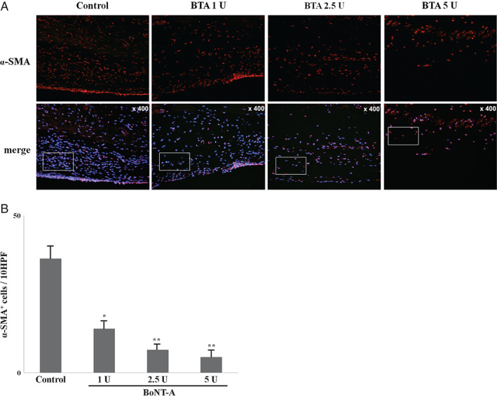
Figure 2.

Immunofluorescence microscopic sections of capsular tissues in experimental groups and control group at the 30‐day time point (original magnification, × 400). To quantify the myofibroblasts, sectioned tissues were immunostained with anti‐alpha‐smooth muscle actin (α‐SMA) antibody and the α‐SMA + cells were counted in four randomly selected fields. The number of α‐SMA + cells in capsules was decreased in experimental groups compared with control group (P < 0·05).*P < 0·05 versus control; **P < 0·01 versus control.
