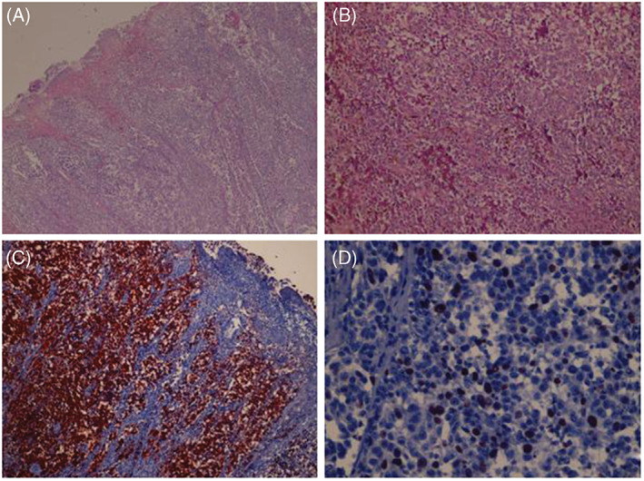
Figure 3.

(A) Nodular tumoural lesion causing destruction of the surface epithelium and ulceration (H & E, ×50). (B) Closer view shows large neoplastic cells with prominent nucleoli and brownish cytoplasmic melanin pigment (H&E, ×200). (C) Neoplastic cells reveal diffuse HMB45 positivity (immunoperoxidase, HMB45, ×200). (D) 20% of neoplastic cells demonstrate Ki‐67 positivity (immunoperoxidase, Ki‐67, ×200).
