Figure 2.

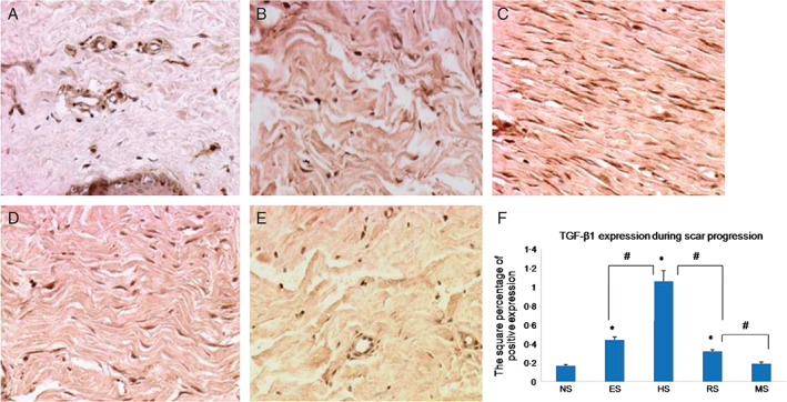
Immunohistochemical staining with transforming growth factor (TGF)‐β1 antibodies in normal skin (A), early scars (B), proliferative scars (C), regressive scars (D) and mature scars (E). Positive cells are indicated in brown colour. (F) The quantification of the microvessel density at each scar stage (n = 8; original magnification, 200×). * indicates a significant difference compared to the normal control (P< 0·05); # indicates a significant difference compared to the former stage (P< 0·05). (NS, ES, HS, RS and MS represent normal skin, early scars, proliferative scars, regressive scars and mature scars, respectively).
