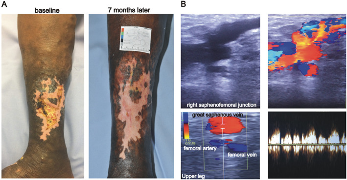
Figure 1.

A leg ulcer with pulsating varicose veins. (A) The patient has a lower leg ulcer of about 4 cm in diameter at the right medial calf. Please note the varicose veins above the ulcer. Healing of the wound was achieved within 7 months after presentation of the patient. (B) Duplex sonography reveals incontinence of the saphenofemoral junction with reversed arterial‐like pulsating flow during the whole course of the great saphenous vein. At the upper leg, both the femoral vein and great saphenous veins show reversed arterial‐like pulsating flow. The Duplex image displays phasic wave forms in the great saphenous vein of the upper leg.
