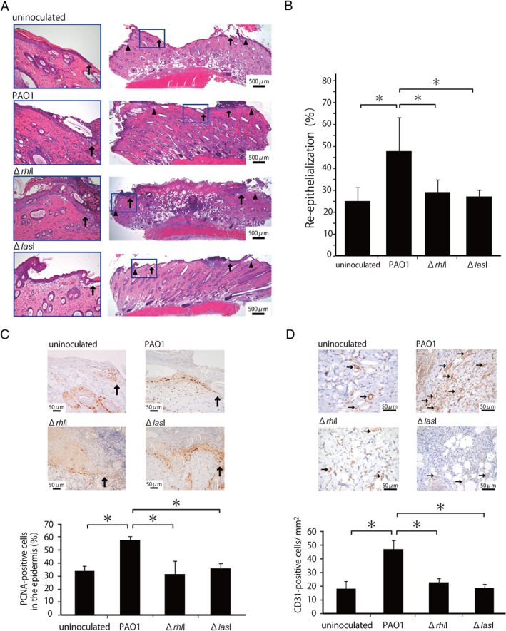
Figure 1.

Effect of the rhlI‐defect on skin wound healing promoted by Pseudomonas aeruginosa. Wounds were created and a suspension of PAO1 or ΔrhlI or ΔlasI was inoculated into the wounds. Uninoculated wounds were created in other rats. (A) Representative histological views of skin wounds on day 3. Arrowheads and arrows indicate the original wound edges and re‐epithelialised leading edges, respectively. (B) Re‐epithelialisation extent was calculated on day 3 (n = 6). (C) Cellular proliferation in the epidermis at the wound edges. Arrows indicate proliferating cell nuclear antigen (PCNA)‐positive microvessels. The percentages of PCNA‐positive cells among the 500 cells in the epidermis on day 3 (n = 3). (D) Microvessel counts on day 3. Arrows indicate CD31‐positive microvessels. The vascular density/mm2 was determined by counting the positive vessels within six visual fields (n = 3). Each column represents the mean ± standard deviation (SD). *P < 0·05.
