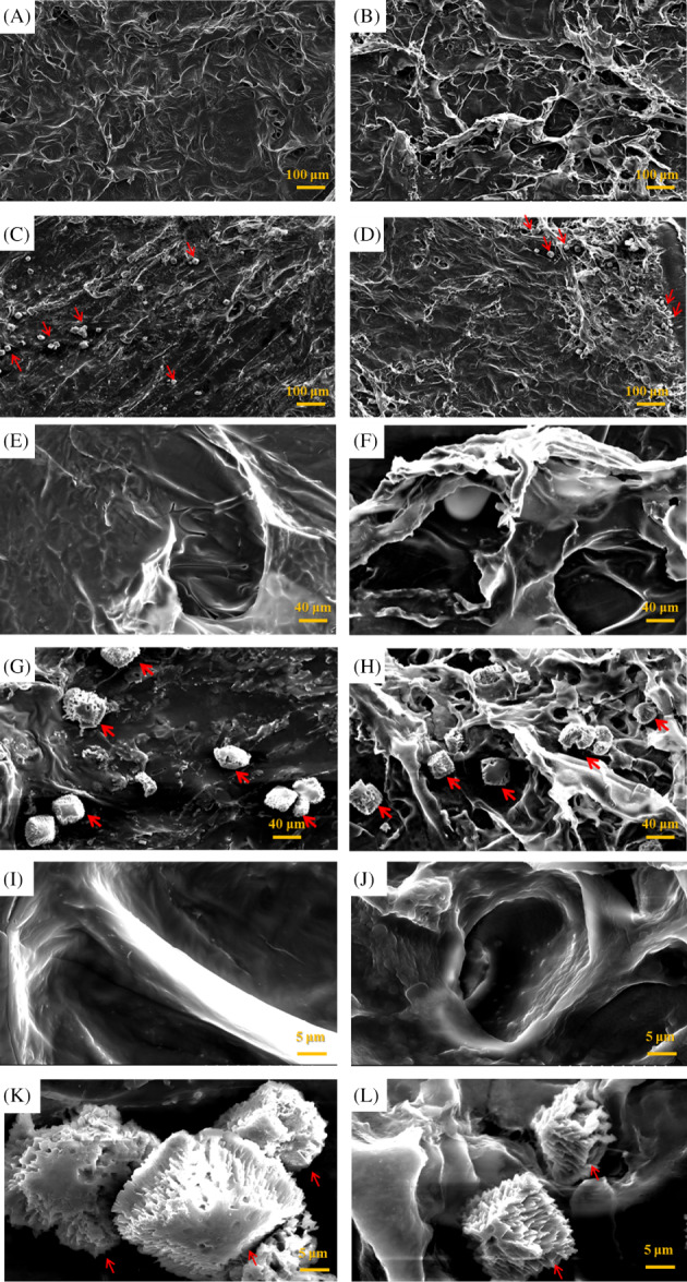
Figure 1.

Scanning electron micrographs of a collagen scaffold (A, E, I), human adipose stem cells (hASCs) seeded on a collagen scaffolds (C, G, K), a collagen/RSV scaffolds (B, F, J), and hASCs seeded on a collagen/RSV scaffolds (D, H, L). Red arrows indicate cells. The surface of the collagen/RSV scaffolds is rougher and more creased than that of the collagen scaffolds. Both scaffolds exhibit excellent cell compatibility. Scale bar = 100 μm (A–D); 20 μm (E–H); 5 μm (I–L)
