
Figure 4.
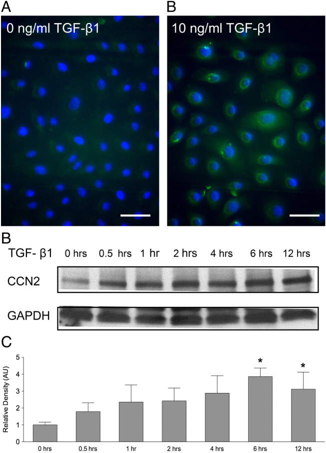
Transforming growth factor β1 (TGF‐β1) increases connective tissue growth factor (CCN2) protein production in keratinocytes. CCN2 protein production in vitro was detected by immunohistochemistry. Green staining indicates CCN2, and blue staining indicates nuclei. (A) After 24 hours' culture in a serum‐free control culture media, keratinocytes expressed low levels of CCN2. (B) The addition of 10 ng/ml TGF‐β1 increased CCN2 protein production. The experiments were repeated three times with similar results. Scale bar equals 20 µm. (C) TGF‐β1‐induced CCN2 protein production was detected with western blot. An increased production of CCN2 was observed after the addition of TGF‐β1, and significantly increased levels of CCN2 were detected after 6 and 12 hours. One representative western blot out of three independently performed experiments is shown. (D) The relative density of the bands was normalised to GAPDH. Graphs show mean ± SD from three independent experiments, n = 6. *P < 0·05 versus untreated control.