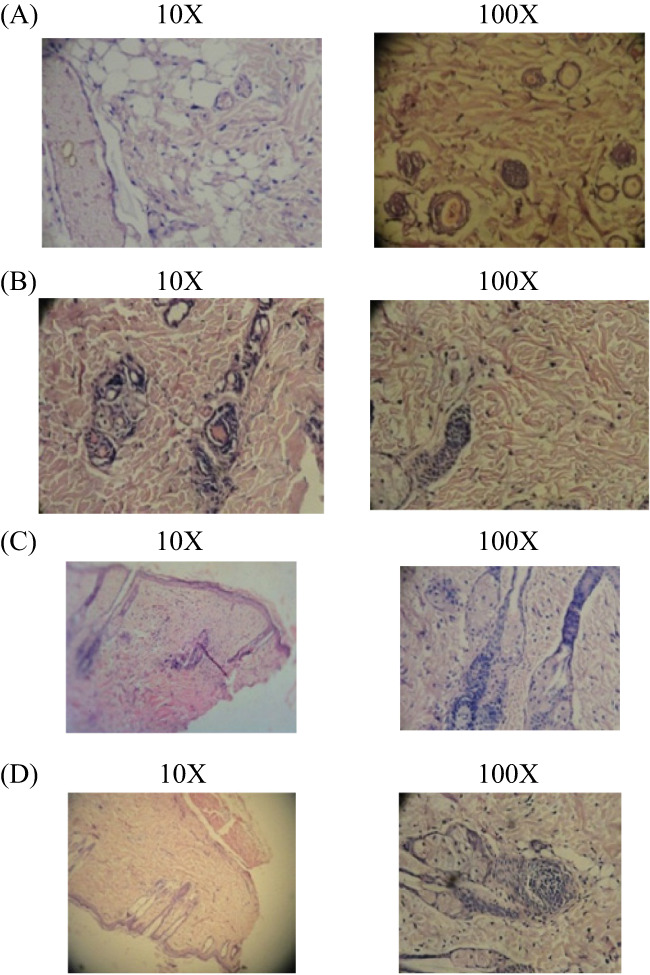
Figure 7.

(A‐D) Histopathology study of excision wound healing. In the excision wound model, 4 groups are present, the results of histopathological changes are presented. (A) Group I: vehicle control (ointment base)—collagen not fully formed, scanty granulation tissue with minimum fibroblast; (B) Group II: standard control—better granulation tissue formation and collagenisation with fibroblast; (C) Group III: Cb‐peptide ointment (low dose)—greater degree of granulation tissue formation and collagenisation with fibroblast; (D) Group IV: Cb‐peptide ointment (high dose)—maximum collagenisation, epithelialisation was early and complete
