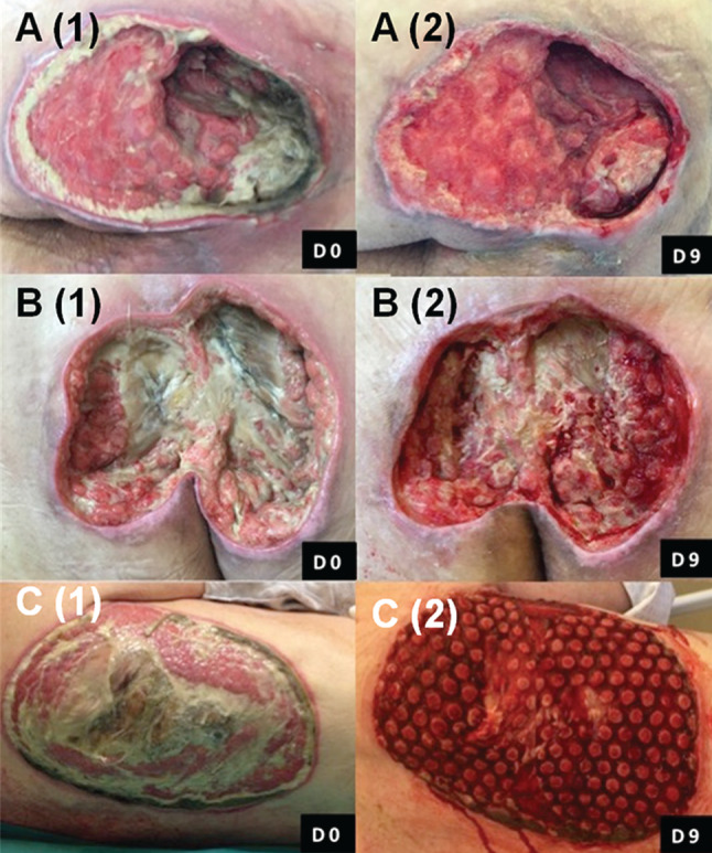
Figure 3.

Wound‐healing progressions of three different pressure ulcers (A–C) in this series. Each pressure ulcer, located in the perineal region, is shown at Day 0 and Day 9 after three successive applications (9 days) of NPWTi‐d with ROCF‐CC. Reduction of fibrinous tissue and cleansing of the wound as well as granulation tissue formation were noted at each dressing change.
