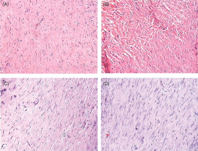
Figure 2.

Area corresponding to cutaneous injury that shows fibroplasia. (A) Control group, 21 days after cutaneous injury. (B) Laser group, 21 days. (C) Control group, 35 days after cutaneous injury. (D) Laser group, 35 days (Haematoxylin‐eosin, 400×)
