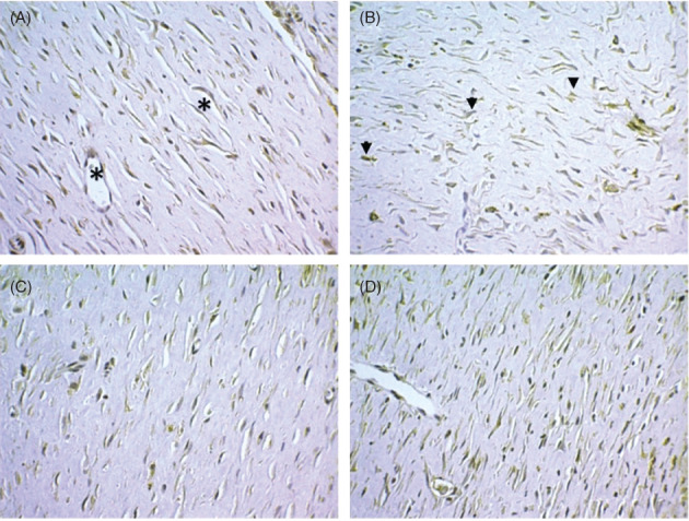
Figure 4.

Expression of VEGF+ cells in the area corresponding to fibroplasia. (A) In the outline of small capillaries (asterisks). (B) As isolated entities dispersed in the conjunctive matrix (arrows). (C) Control group, 21 days after the cutaneous wound was made. (D) Laser group, 21 days. (Immunohistochemistry, anti‐VEGF, 400×)
