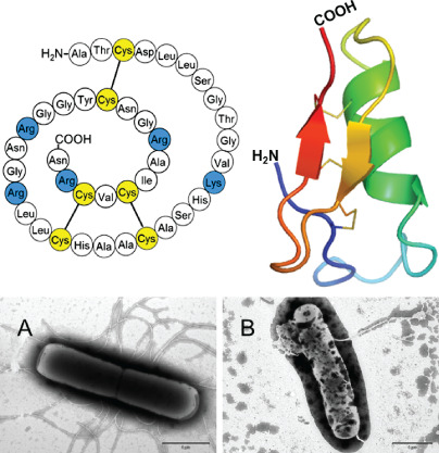
Figure 3.

The amino acid sequence of lucifensin (Lucilia sericata defensin), its tertiary structure and its effect on bacterial cell walls. Electron micrographs of negatively stained Bacillus subtilis either untreated (A) or treated by lucifensin for 60 minutes (B). Scale bars represent 1 µm. Illustrated representation of the three‐dimensional structure of lucifensin (PDB ID: 2LLD) generated by Pymol (http://www.pymol.org).
