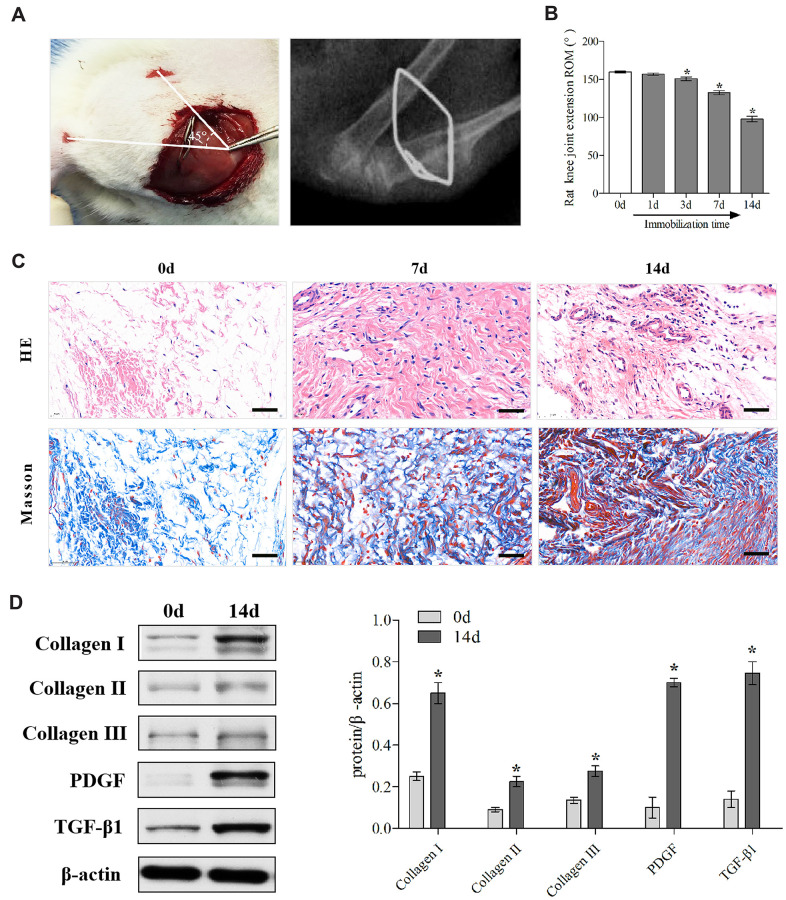
Figure 1.
Establishment and identification of the rat knee joint PTJC model. (A) Schematic and X-ray of rat knee joint post-traumatic immobilization. (B) Measurement of extension ROM of the affected knee joint at Day 0, 1, 3, 7, and 14 post-modelling. (C) HE and Masson staining of the posterior joint capsule of the affected knee at day 0, 7, and 14 post-modelling. Scale bars, 50 μm. (D) Expression of fibrosis-associated protein (collagen I, collagen II, collagen III, PDGF, and TGF-β1) in the posterior joint capsule at day 0 and 14 post-modelling were assessed via western blot. Endogenous β-actin was used as an internal control. Error bars represent standard deviation. *P <0.05 compared with the day 0 group.

