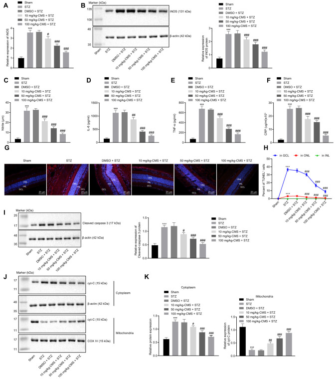
Figure 3.
CMS reduced oxidative stress, inflammation, and apoptosis in DR rats. STZ-treated rats were treated with DMSO, 10 mg/kg CMS, 50 mg/kg CMS, and 100 mg/kg CMS. n = 15 per treatment. (A) Expression pattern of iNOS as determined by RT-qPCR in rat retinal tissues, normalized to β-actin. (B) Representative Western blots of iNOS protein and its quantitation in rat retinal tissues, normalized to β-actin. (C) Expression of NO measured by nitrite test in rat retinal tissues. (D–F) Expression patterns of IL-6 (D), TNF-α (E), and CRP (F) measured by ELISA in the cell supernatant. (G, H) Representative images of apoptotic cells (× 400) (scale bar = 25 μm) (G) and cell apoptosis in rat retinal tissues (H) by TUNEL. (I) Representative Western blots of cleaved caspase-3 protein and its quantitation in rat retinal tissues, normalized to β-actin. (J, K) Representative Western blots of Cyt-C protein and its quantitation in the cytoplasm and mitochondria, normalized to β-actin. * p < 0.05, ** p < 0.01, *** p < 0.001, compared to the sham-operated rats, and # p < 0.05, ## p < 0.01, ### p < 0.001, compared to the rats injected with STZ and treated with DMSO. The results were measurement data, which were expressed as mean ± standard deviation. Comparisons between multiple groups were analyzed by one-way ANOVA with Tukey’s post hoc test (n = 15). iNOS, inducible nitric oxide synthase; CMS, coumestrol, STZ, streptozotocin; DMSO, dimethyl sulfoxide; NO, nitric oxide; RT-qPCR, reverse transcription-quantitative polymerase chain reaction; IL-6, interleukin-6; TNF-α, tumor necrosis factor α; CRP, C-reactive protein; ELISA, Enzyme linked immunosorbent assay; GCL, ganglion cell layer; INL, inner nuclear layer; ONL, outer nuclear layer; Cyt-C, cytochrome c; ANOVA, analysis of variance; n, number.

