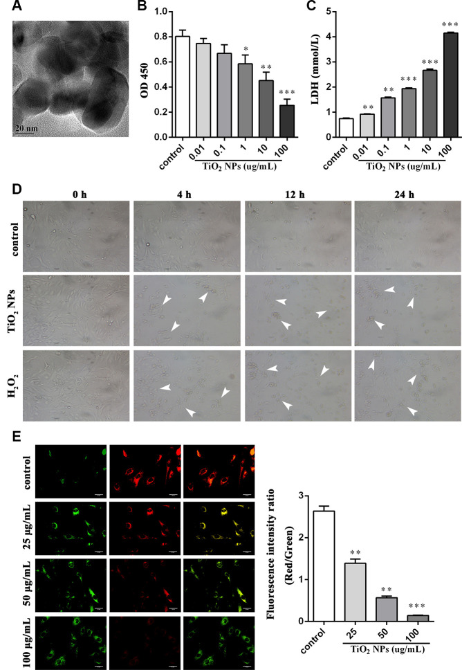
Figure 1.
TiO2 NPs boosted cell damage of corneal endothelial cells. (A) Detection of shape and size of TiO2 NPs by TEM, bar = 20 nm. (B) Cell viability of TiO2 NPs-treated primary endothelial cells. (C) Measurement of LDH content in TiO2 NPs-treated primary endothelial cells. (D) Morphological analysis of TiO2 NPs-treated primary endothelial cells by inverted microscope, bar = 50 μm. (E) Mitochondrial membrane potential of TiO2 NPs-treated primary endothelial cells by using JC-1, bar = 30 μm. *P<0.05, **P<0.01, ***P<0.001.

