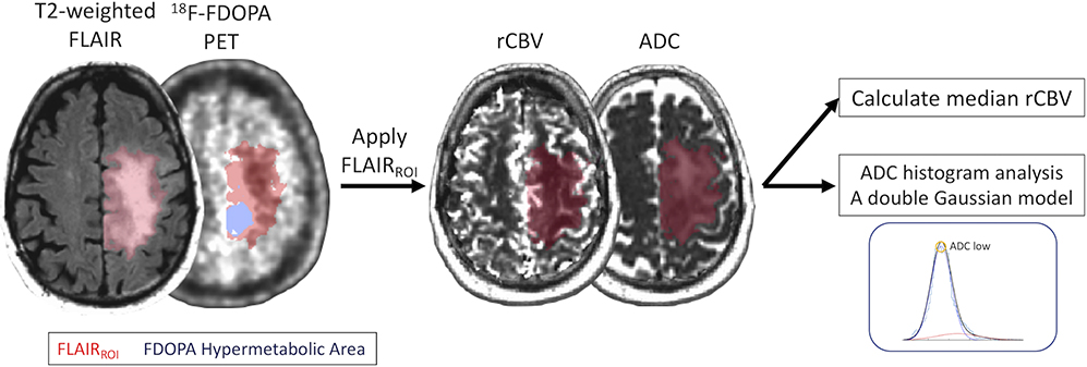
Figure 1.
Postprocessing and segmentation example. A 57-year-old man with newly diagnosed astrocytic diffuse glioma (WHO grade II, IDH1 mutant, 1p19q non-codeleted, and MGMT methylated status). ROIs of the FLAIR hyperintense region (FLAIRROI, red area) and 18F-FDOPA hypermetabolic area (nSUV > 1, blue area) within FLAIRROI are shown. nSUVmax and volumes for each ROI are calculated. FLAIRROI is copied and pasted on rCBV and ADC maps, and median rCBV and ADClow are calculated.

