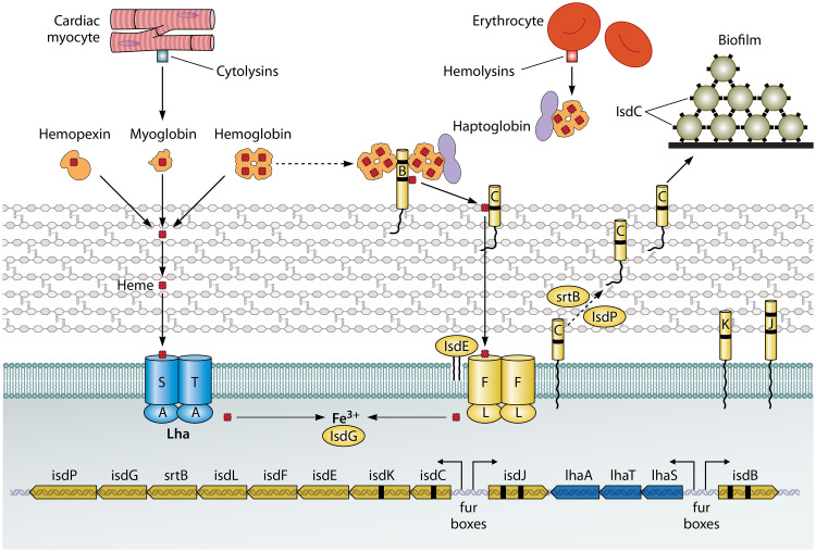
FIG 1.
Iron acquisition from heme. The lower part of the diagram shows the genetic organization of the isd locus of S. lugdunensis. The locations of fur boxes and the directions of transcription following induction by iron limitation are shown by arrows. The upper part of the diagram shows the cell membrane and cell wall peptidoglycan and the locations of the Isd proteins (right side) and the Lha proteins (left side). Hemoproteins are shown with their bound heme molecules (red rectangles), including four in hemoglobin and one in myoglobin and hemopexin. Myoglobin and hemoglobin are released from myocytes and erythrocytes, respectively, by the action of cytolytic toxins. Black dashes in genes and proteins indicate NEAT domains. IsdB NEAT1 can bind hemoglobin and the haptoglobin-hemoglobin complex. The passage of heme across the cell wall and membrane is shown, along with release of Fe3+ by intracellular hemoxygenase. The predicted second hemoxygenase is not shown. IsdC proteins on different cells also promote biofilm formation.

