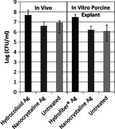
Figure 6.

Comparison of Pseudomonas aeruginosa biofilm cultured on an ex vivo porcine skin partial‐thickness ‘wound’ explants to P. aeruginosa biofilm cultured on an in vivo pig burn model after 24‐hour exposure to silver dressings: Hydrocolloid–Ag (Contreet‐H®‐Ag), Hydrofibre–Ag (Aquacel®‐Ag) and Nanocrystalline–Ag (Acticoat®). No statistically significant difference was found between any dressing treatments.
