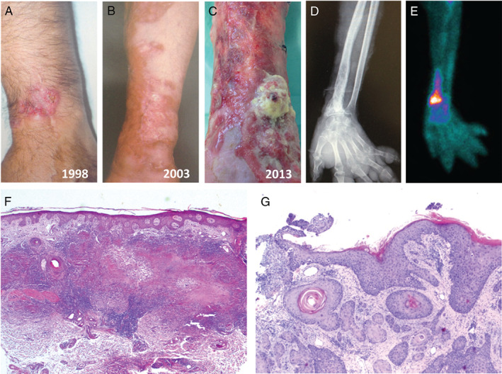
Figure 1.

(A) Erythematous round plaque with yellow atrophic centre on the left wrist. (B) Erythematous brown plaque of necrobiosis lipoidica (NL) – centrifuge growth. (C) Friable vegetating tumour lesion on the distal radial area of the left forearm. (D) Digital radiography of the left forearm and hand: honey‐comb pattern in the distal radius. (E) Bone scintigraphy with Technetium‐99m, increased fixation of the isotope in the distal radius. (F) Haematoxylin and eosin (H&E), ×25 NL. (G) H&E, ×40 squamous cell carcinoma.
