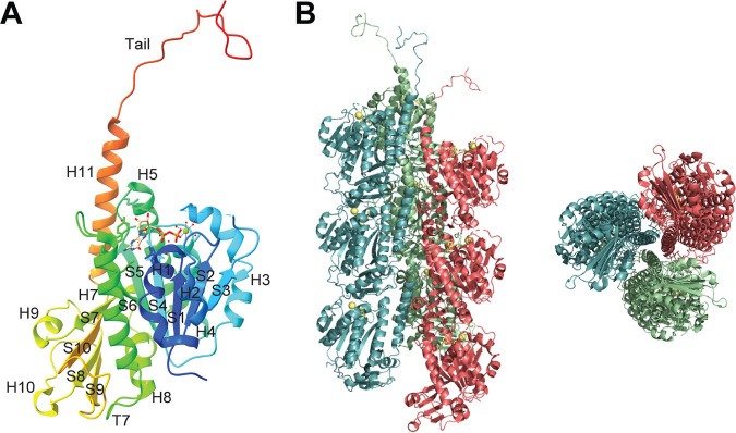
FIG 1.
Monomeric and filament crystal structures of phage 201Ф2-1 PhuZ. (A) Cartoon representation of the crystal structure of a PhuZ201 monomer annotated with secondary structural elements (PDB ID 3R4V) (53). The bound GDP-Mg2+ is shown in ball-and-stick format. (B) Cartoon representation of the crystal structure of PhuZ201 filament, with individual protofilaments presented in different colors (PDB ID 3J5V) (67). The bound GDP-Mg2+ elements are shown in ball-and-stick format. (Left) Side view; (right) en d-on view.

