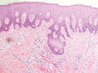
Figure 2.

Histological examination of the skin lesions in a patient with hidradenitis suppurativa. The examination showed the presence of epidermal hyperplasia, dermal infiltration of inflammatory cells, multinucleated giant cells, granulomatous changes and a small abscess formation (HE staining, ×40).
