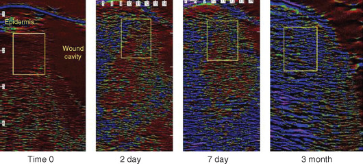
Figure 2.

Changes in levels of oedema in periwound tissue of pressure ulcers during gauze‐based negative pressure wound therapy (NPWT) at−80 mmHg. The region of interest where the pixel analysis was carried out is indicated by the boxed area on each image. The region of interest was placed in the same location on each scan for time 0, day 2, day 7 and 3 months.
