
Figure 4.
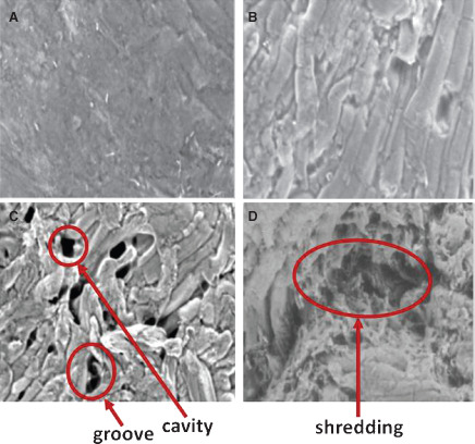
Scanning electron microscope images of ex vivo model wound surfaces after cleansing treatments. Areas of trauma and disruption are indicated by arrows. (A) No treatment at 500× magnification. (B) Negative pressure wound therapy with instillation (NPWTi) at 402× magnification. (C) Low‐pressure lavage at 332× magnification. (D) High‐pressure lavage at 395× magnification.