
Figure 4.
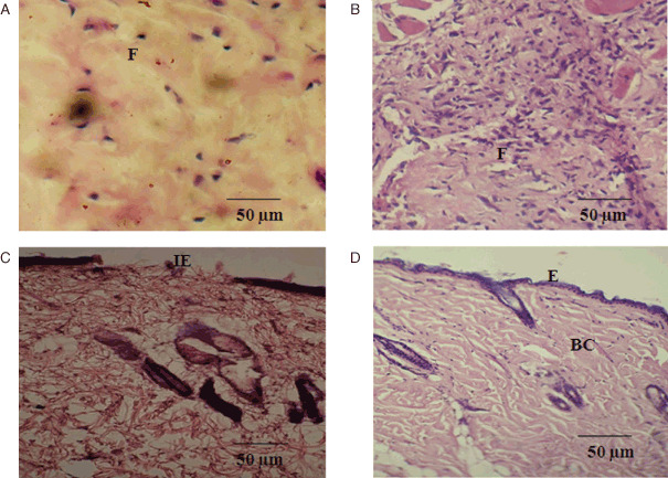
Photomicrographs of histological study of control and Annona squamosa treated wound tissues on weeks 1 and 2 of post‐wounding, respectively (stained with haematoxylin–eosin and Van Gieson's; magnification 20×). During week 1, control (A) showing loosely packed collagen with irregular epithelialisation and less fibroblasts treated tissue (B) showing new blood vessel formation and high fibroblasts with dense collagen deposition. On week 2, control (C) shows thin epithelial layer with less collagen and treated (D) shows complete epithelialisation with regularly arranged dense collagen. Scale bar 50 µm. BC, blood capillaries; E, epithelialisation; F, fibroblast; IE, incomplete epithelialisation.