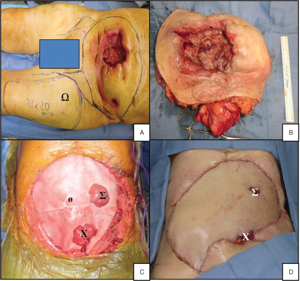
Figure 3.

Intraoperative photographs. (A) Plot of the resection area and free flap harvesting zone (Ω). (B) En bloc resection of the abdominal wall and the small intestine. (C) Implementation of bioprostheses (ø) with one ileostomy (Σ) and one colostomy (X) through the prostheses. (D) Local view at the end of surgery.
