
Figure 2.
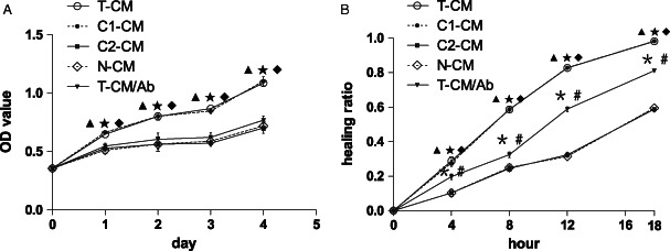
Function of BMSCs expressing exogenous hVEGF165/hBD3. (A) Proliferation‐promoting effect of different CMs. The cell growth curves revealed that T‐CM and C1‐CM significantly promoted cell proliferation from days 1 to 4, compared with C2‐CM, N‐CM and T‐CM/Ab. The addition of anti‐hVEGF165 neutralising antibody to T‐CM inhibited cell growth significantly. (B) In vitro wound‐healing assay on monolayer of cells. The HR was significantly higher for cells treated with T‐CM and C1‐CM at 4, 8, 12 and 18 hours. The anti‐hVEGF165 neutralising antibody (T‐CM/Ab) significantly inhibited T‐CM‐stimulated wound healing, but still did not abolish the effect of T‐CM on wound healing. The HR was significantly higher for T‐CM/Ab‐treated cells than C2‐CM‐ and N‐CM‐treated cells at 4, 8, 12 and 18 hours. No significant differences were found between T‐CM and C1‐CM and between C2‐CM and N‐CM.  P < 0·05 T‐CM or C1‐CM versus C2‐CM,
  P < 0·05 T‐CM or C1‐CM versus N‐CM,
  P < 0·05 T‐CM or C1‐CM versus T‐CM/Ab,
  P < 0·05 T‐CM/Ab versus C2‐CM,
  P < 0·05 T‐CM/Ab versus N‐CM.