
Figure 5.
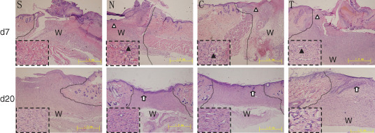
Histological studies on haematoxylin–eosin‐stained radiation‐wound tissues. The amplified local areas are shown in the dashed boxes. The comparable image of each group includes the wound edge and frontier of migrated epithelial cells. Upper panels: on day 7 after injury, blood congestion and haemorrhage were apparent in group S, mild in groups C and N and invisible in group T. Meanwhile, actively proliferating epithelial cells formed a multilayer structure (white triangle) and migrated to the centre of the nude wound. Angiogenesis in granulation tissues (black triangles) was clear in group T, mild in groups C and N and invisible in group S. Lower panels: on day 20 after injury and treatment, the wound was re‐epithelialised completely with regenerated skin appendages (white arrows) in group T, but the re‐epithelialisation and regeneration of skin appendages were less in groups C and N and invisible in group S. The lines indicate the interface of the wound and unaffected skin. W, wound site. Bars = 1 mm.