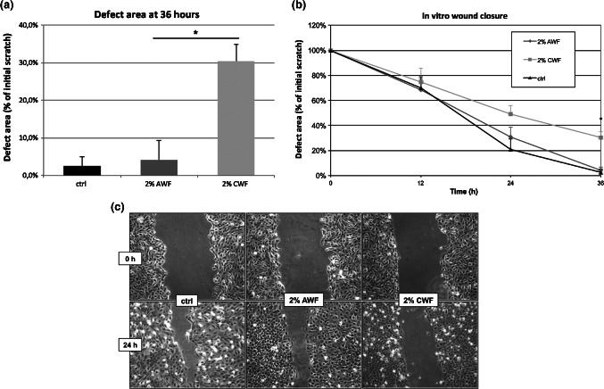
Figure 2.

Scratch assay. Keratinocytes were plated onto microplates. After 24 hours, they were incubated with 10 µg/ml mitomycin C for 2 hours to inhibit cell proliferation. The monolayer was scratched with a plastic pipette tip. Culture medium was changed (black) or replaced by 2% acute wound fluid (dark grey) or 2% chronic wound fluid (light grey) and in vitro epithelialisation was documented. Wound closure was evaluated by measuring the remaining cell‐free area and expressed as percentage of the initial cell‐free zone. The results of three independent experiments as mean ± SD are shown. *P ≤ 0·05. (A) Remaining cell‐free area after 36 hours. (B) Time course of epithelialisation. (C) Representative photo documentation at 24 hours.
