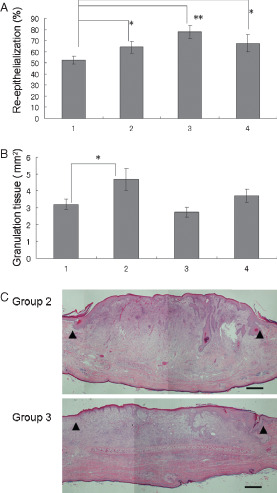
Figure 5.

Assessment of topical reagents for skin wounds. (A) Reepithelialization; (B) area of granulation tissue. Lane 1: control (dressing only), n = 11; lane 2: application of basic fibroblast growth factor (bFGF) spray, n = 14; lane 3: application of prostaglandin E1 (PGE1) ointment, n = 12; lane 4: application of dibutyryl cyclic adenosine monophosphate ointment, n = 11. *P < 0·05, **P < 0·01. The data obtained were analysed by unpaired Student's t‐test. Each bar represents mean ± standard error of the mean. (C) Representative histopathology of wounds applied with topical reagents. Group 2: a wound treated with bFGF spray; group 3: a wound treated with PGE1 ointment. Arrow heads indicate the original wound edges (haematoxylin and eosin staining, scale bar: 500 µm).
