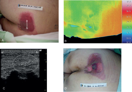
Figure 3.

Case 14. (A) A 50‐year‐old male had a d2 pressure ulcer on his sacral region. The bar indicates the location of the ultrasonography probe. (B) The thermographic findings showed that the wound temperature was relatively high and (C) ultrasonography showed an unclear layered structure and a heterogeneous hypoechoic area (white dashed circle) at the primary examination. (D) The lesion worsened and was estimated as stage DU (stage D3 or worse) within 1 week, indicating a deep tissue injury. Line a–b indicates the direction of the applied probe in panels (A) and (C).
