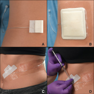
Figure 1.

Photo sequence showing: (A) an artificial wound model (AWM) placed on the lower left back of a volunteer; (B) placement of a foam dressing over the AWM; (C) securement of the catheter and hub to the front of the volunteer for easy access and (D) injection of artificial wound fluid into the wound model. Injection of the model can be done by the study coordinator (as pictured) or by the subject in between assessment visits.
