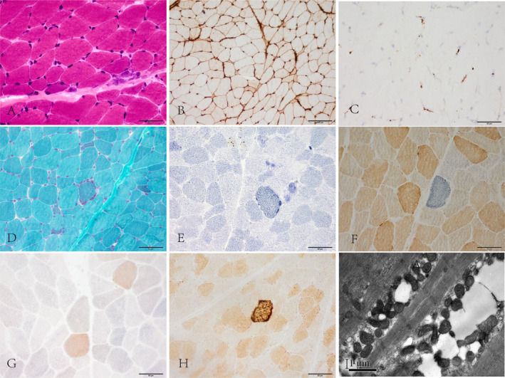
Figure 1.

(A) Scattered atrophy, regenerated muscle fibers. HE staining. (B) Diffuse sarcolemmal MHC‐I positive immunoreactivity. (C) Mild membrane attack complex deposition on endomysial capillaries. (D) Atypical ragged red fibers appeared by modified Gomori trichrome staining. (E) SDH hyper‐reactive fibers. (F) COX‐negative fibers. (G) Numerous COX‐deficient fibers, not restricted to the perifascicular region. SDH‐COX co‐staining. (H) COX hyper‐reactive fibers. Original magnification, ×40. (I) Mitochondrial proliferation and abnormal aggregation among myofibrils. Electron microscopy. Scale bar are 1 μm. MHC‐I, major histocompatibility complex class‐I; COX, cytochrome oxidase C; SDH, succinate dehydrogenase.
