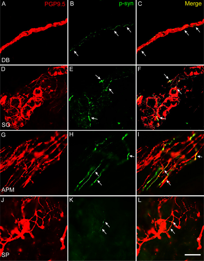
Figure 3.

P‐syn deposited in skin nerve fibers of PD patients with and without G2385R variant. Double immunofluorescent staining demonstrated linear or dotted p‐syn immunosignals (green) colocalized with PGP9.5 signals in DB in deep dermis (A–C), nerve fibers innervating SG (D–F), APM (G–I), and SP and DB (asterisk) in superficial dermis (J–L). P‐syn, phosphoralated α synuclein; Arrows indicated p‐syn immunosignals; DB, dermal nerve bundles, SG, nerve fibers innervating sweat glands, APM, arrector pili muscles; SP, subepidermal plexus; PD, Parkinson disease. Scale bar: 50 μm.
