
Figure 5.
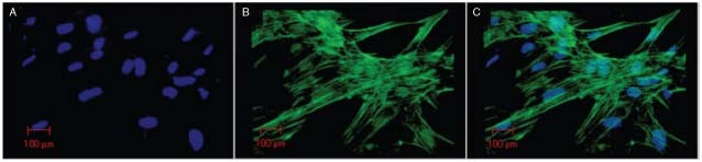
Confocal microscopy. Cultured fibroblasts from the adjacent skin. (A) Cell nuclei stained with DAPI (blue); (B) actin filaments immunostained with phalloidin/AlexaFluor‐488 (green); (C) overlapped images.
Official websites use .gov
A
.gov website belongs to an official
government organization in the United States.
Secure .gov websites use HTTPS
A lock (
) or https:// means you've safely
connected to the .gov website. Share sensitive
information only on official, secure websites.
Confocal microscopy. Cultured fibroblasts from the adjacent skin. (A) Cell nuclei stained with DAPI (blue); (B) actin filaments immunostained with phalloidin/AlexaFluor‐488 (green); (C) overlapped images.