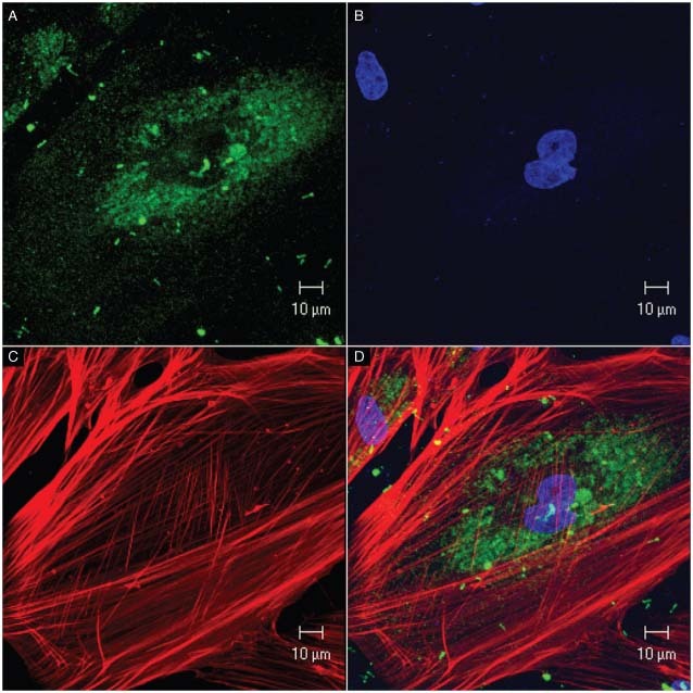
Figure 6.

Confocal microscopy. Cultured fibroblasts from the adjacent skin. (A) Mitochondria stained with MitoTracker Green; (B) cell nuclei stained with DAPI (blue); (C) actin filaments immunostained with phalloidin/AlexaFluor‐594 (red); (D) overlapped images.
