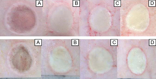
Figure 1.

Macroscopic findings after wounding (A), and on day 1 (B), day 2 (C) and day 3 (D) after loading in the 10‐kg (upper) and 20‐kg (lower) groups using images from the same rat for each group. After pressure unloading, a dark red circle with oedema at the circumference was seen in the compressed areas in both 10‐kg and 20‐kg loading groups. In both groups, the tone colour of the wound surface area turns yellowish white on days 1, 2 and 3. There are no differences in the macroscopic appearances between the 10‐kg and 20‐kg loading groups.
