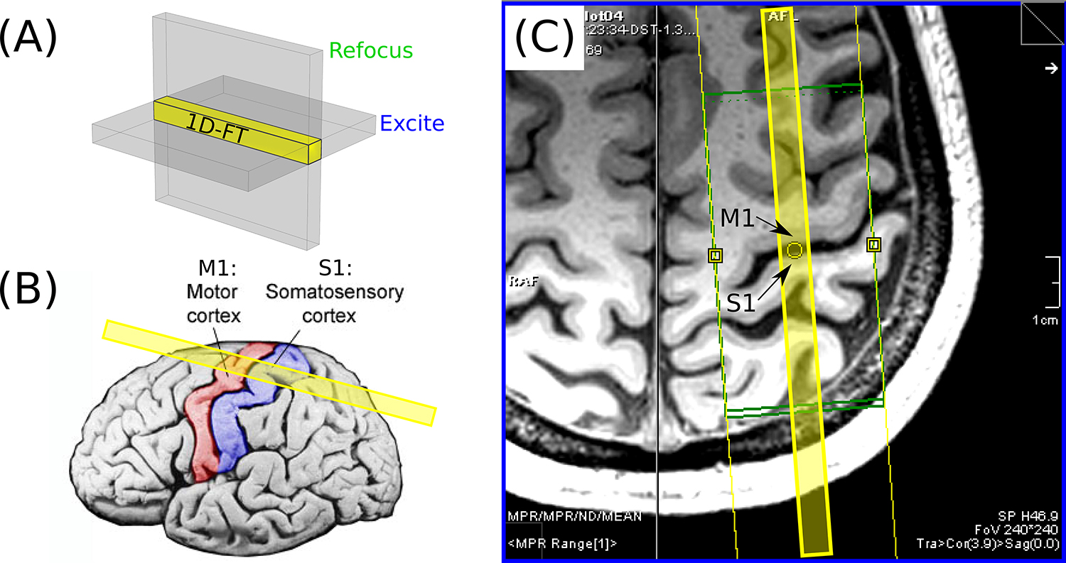
FIG. 2.

(A) An illustration of the concept underlying the spin-echo line-scan technique (2–4): by applying the slice-select gradients for the RF excitation and refocusing pulses orthogonally to one another, the MR signal arises from the column of spins within the intersection of the two slices, shown in yellow. This “line” only requires Fourier encoding in one direction and can thus be reconstructed via a 1D Fourier Transform (FT), with no need for phase encoding. (B) The prescription of the line shown with respect to a surface rendering of cortex, with M1 and S1 colored red and blue, respectively. (C) A slice through the 3D MPRAGE data of volunteer 1, as seen on the scanner console. Note the relatively flat patch of M1 and S1 adjacent to the yellow circle; the line (shown in yellow overlay) was prescribed to be as perpendicular as possible to cortex at this location.
