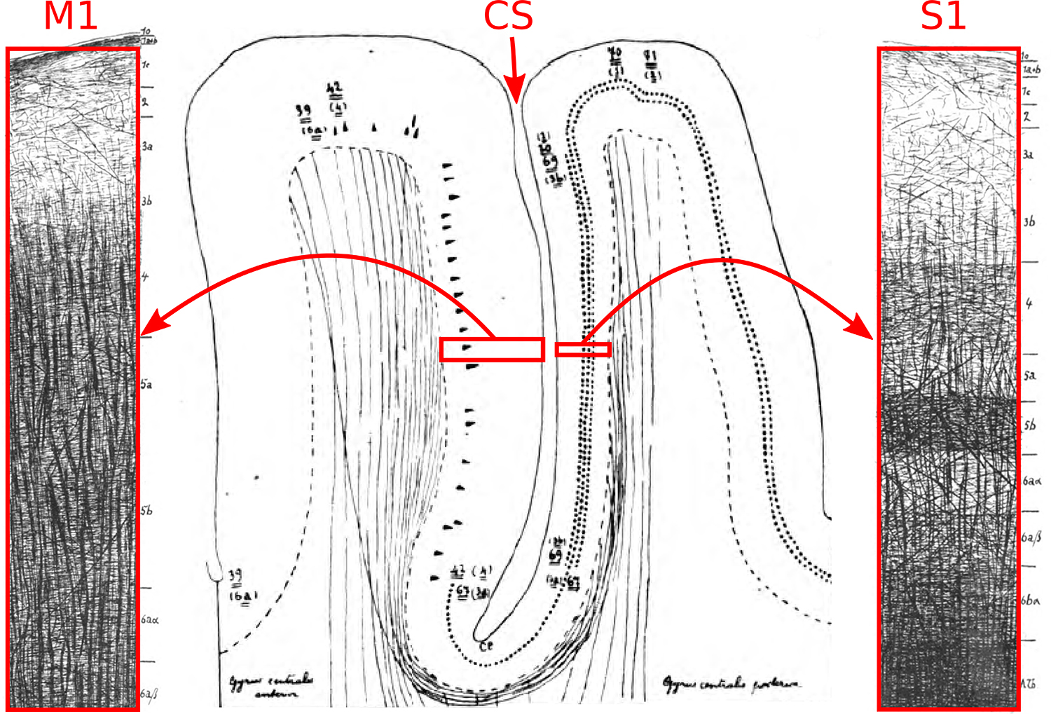
FIG. 8.

Myeloarchitecture of M1 and S1, adapted from the seminal monograph of Vogt and Vogt (9). In M1, note the dominance of radial myelinated fibers at most cortical depths, and in S1, note the pronounced tangential myelinated fibers in the deep layers. CS=central sulcus.
