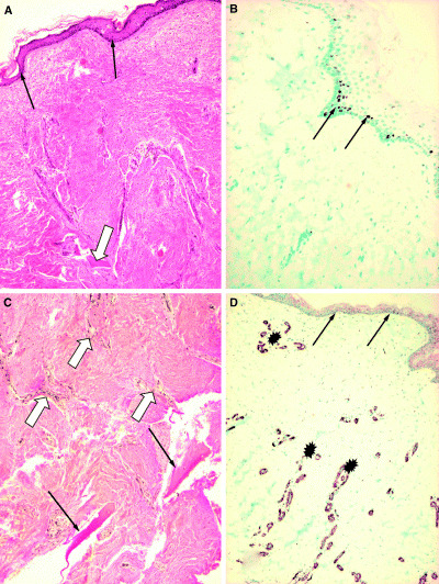
Figure 3.

Punch biopsy specimen taken from the site of the acellular dermal matrix implant 2.5 years after grafting. Histopathological evaluation: (A) Hematoxilin‐eosin stain of the graft site shows atrophic but intact and inflammation‐free skin with normal epidermal structure and keratinisation. The arrows indicate the intact epidermal layer. The open arrow points to a bony trabecule (original magnification ×10). (B) The basal layer of the epidermis shows normal proliferation capacity of cells, as detected by Ki67 monoclonal antibody (mAb) expression (arrows). The rate of proliferation is comparable with normal epidermis (not shown) (original magnification ×10). (C) Van Gieson staining reveals an oligocellular elastic connective tissue (eosinophilic bundles) in the deep dermis. In the deeper dermal layers, at the osteo‐neo‐dermal junction, mature lamellar bony trabeculae are present (arrows) surrounded by a well‐developed neo‐dermis. Open arrows indicate blood vessels (original magnification ×20). (D) The vasculature of the neo‐dermis at the implant site is shown with mAb to smooth muscle actin in the vessel walls (asterisk) indicating sufficient angiogenesis in the graft during remodelling (original magnification ×10).
