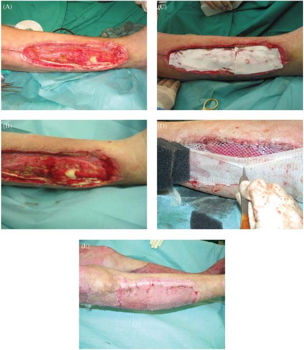
Figure 1.

(A–E) At the lateral part of the right lower extremity an area of about 10 cm2of the extensor‐tendons are exposed (A and B). Single‐stage Matriderm® and STSG 1:1·5 meshed reconstruction (C and D) with a vacuum‐assisted therapy on top of the graft; C shows the Matriderm® on top of the wound, as visible in D after hydration Matriderm® gets transperent, identically to the colour of the wound bed. Wound closure was achieved without regrafting (E).
