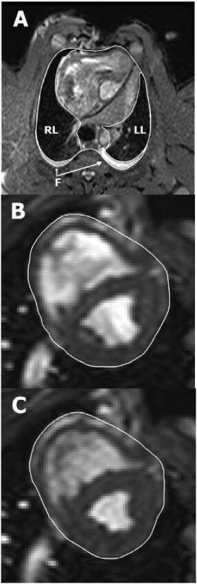
Figure 1.

Illustration of manual delineation of organ volumes. Image (A) shows how the intra‐thoracic structures were outlined in a transverse magnetic resonance (MR) image through the thorax of a pig with an open sternotomy wound. The right lung (RL), left lung (LL) and intra‐thoracic fluid (F) were delineated in each image throughout the entire thorax, allowing the volume of each structure to be quantified. Images (B) and (C) show short‐axis MR images of the same animal's heart in end diastole and end systole, respectively. The pericardial border of the heart was manually delineated in all short‐axis slices covering the entire volume within the pericardium. The total heart volume in end diastole and end systole, as well as the total heart volume variation throughout the cardiac cycle, was then quantified.
