
Figure 2. Caveolae-mediated LDL transcytosis in endothelial cells (ECs) of aortic athero-prone regions.
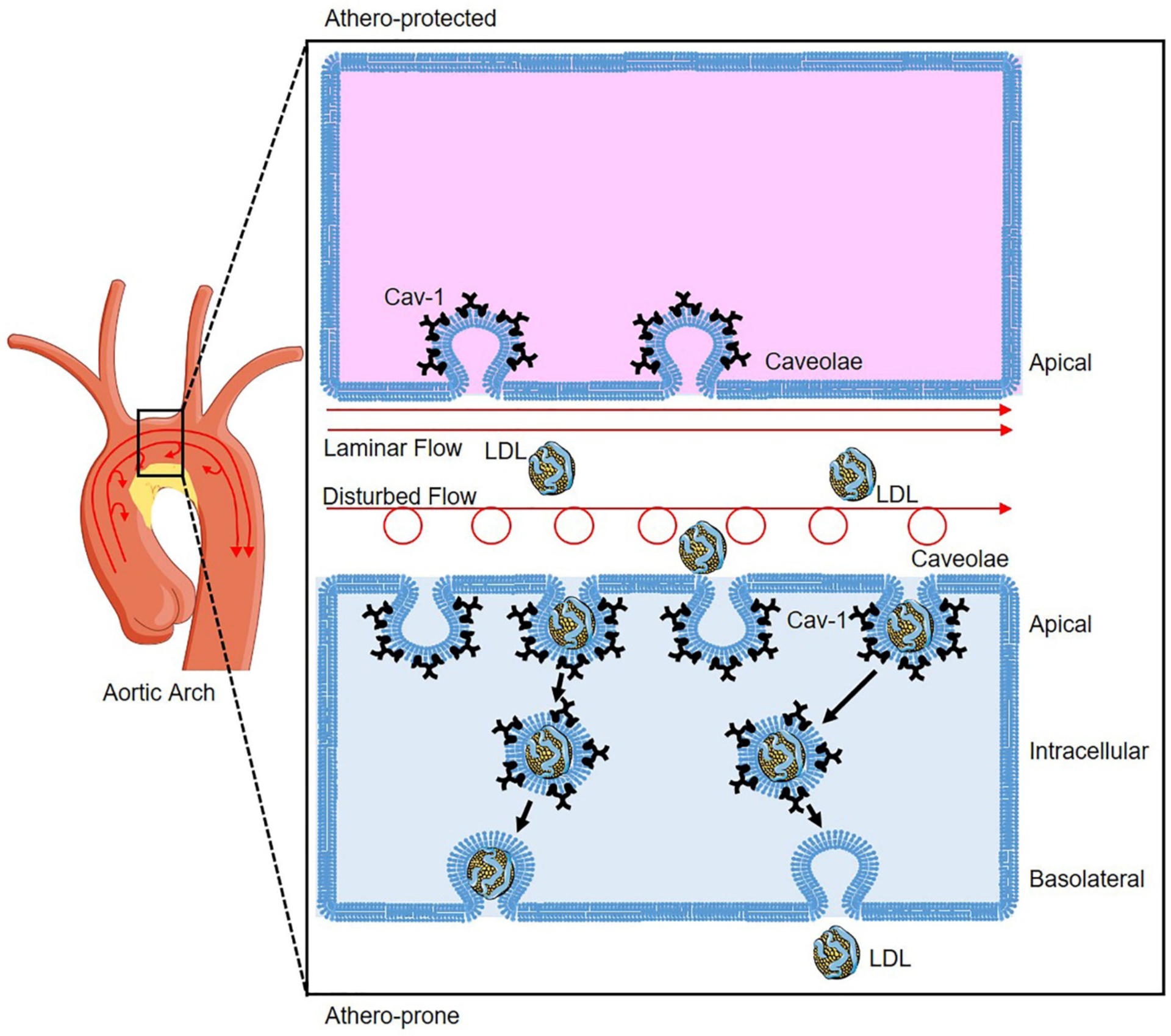
Atherosclerosis occurs preferentially at the athero-prone regions (low curvature) of disturbed flow with low shear stress, whereas laminar flow with high shear stress protects against atherosclerosis. Disturbed flow promotes higher expression of Caveolin-1 (Cav-1) in the athero-prone regions of aortic arch, which induces more caveolae formation compared to ECs of athero-protected regions where laminar flow conditions exist. In the ECs of athero-prone regions, caveolae colocalized with LDL redistribute from apical to intracellular compartment facilitating LDL transcytosis to induce LDL deposition and progression of atherosclerosis.